


VASCULAR ACCESS ULTRASOUND PHANTOM - 4 Vein Bifurcated with Deep Vein
Description
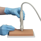
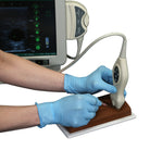
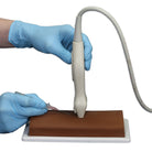
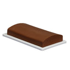
The curved contour on the top of the ultrasound training phantom is similar to contours on patients and has the added benefit of showing the importance of transducer positioning, as positioning can affect the quality of the image viewed–this is not possible on phantoms that are flat. This phantom can be imaged and accessed from the top, side or bottom surface, on the base, or removed from the case.
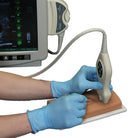
We've been told by clinicians that this ultrasound phantom has the best imaging of all currently available ultrasound training phantoms. Some have even commented that they reduce the gain on the ultrasound machine for advanced users wanting a more challenging ultrasound-guided IV access experience. The ultrasound-guided needle placement is so realistic that movement of the soft tissue can be observed as the needle tip is guided toward the vessel. Successful ultrasound-guided iv insertion can be confirmed by visualization of the needle tip in the vessel and by a blood return.
VATA's Vascular Access Ultrasound Phantom is an excellent model for beginners learning how to start an IV using ultrasound and more advanced clinicians in areas of care including; simulation healthcare labs, IV nursing, anesthesiology, radiology, ultrasound training, nurse practitioners, emergency medicine and others.
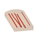
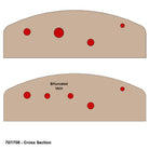
The Vascular Access Ultrasound Phantom contains four vessels:
- Very small superficial vein (3mm vein – 4mm deep)
- Mid-sized vein at medium depth (5mm vein – 7.5mm deep)
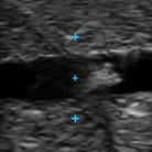
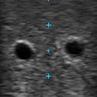
- Mid-sized vein which bifurcates into two smaller vessels (Bifurcated vein 4mm where bifurcated and 7mm when they are one- 12mm deep)
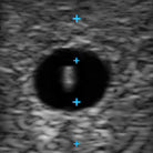
- Larger deeply place vein (7mm vein – 20mm deep)
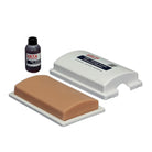
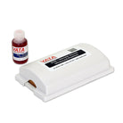
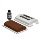
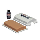
This ultrasound simulation model is supplied pre-filled with a stain resistant simulated blood, a separate 2oz/60ml bottle of stain resistant simulated blood, and a lightweight, compact, form-fitting case that protects the phantom when not in use and is the most compact housing of all currently available phantoms.
Failure to use VATA Inc. ultrasound blood can affect the image quality and will void product warranty. The phantom is compatible with any ultrasound imaging system with an appropriate transducer.
Frequently Asked Questions
How many veins does this phantom have, and how deep are they?
Four: a very small superficial vein (3mm, 4mm deep), a mid-sized vein at medium depth (5mm, 7.5mm deep), a bifurcated vein (4mm per branch, 7mm combined, 12mm deep), and a larger, deeply placed vein (7mm, 20mm deep) — giving learners a realistic range of access difficulty in one model.
Why does the phantom have a curved top instead of a flat surface?
The curved contour matches real patient anatomy and lets learners practice adjusting transducer positioning — a skill that directly affects image quality and is difficult to teach on a flat phantom.
Can I access the phantom from more than one side?
Yes — the phantom can be imaged and accessed from the top, side, or bottom surface, on the base, or removed from its case entirely.
Does this phantom require a specific type of ultrasound blood?
Yes. The phantom should only be used with VATA Inc. ultrasound blood. Using other blood can affect image quality and will void the product warranty.
What's included with the phantom?
The phantom ships pre-filled with stain-resistant simulated blood, plus a separate 2oz/60ml bottle of blood, and a lightweight, compact, form-fitting carrying case.
Who is this phantom designed for?
It's suited to both beginners learning ultrasound-guided IV access and advanced clinicians, and is used across simulation healthcare labs, IV nursing, anesthesiology, radiology, ultrasound training programs, nurse practitioner programs, and emergency medicine.